
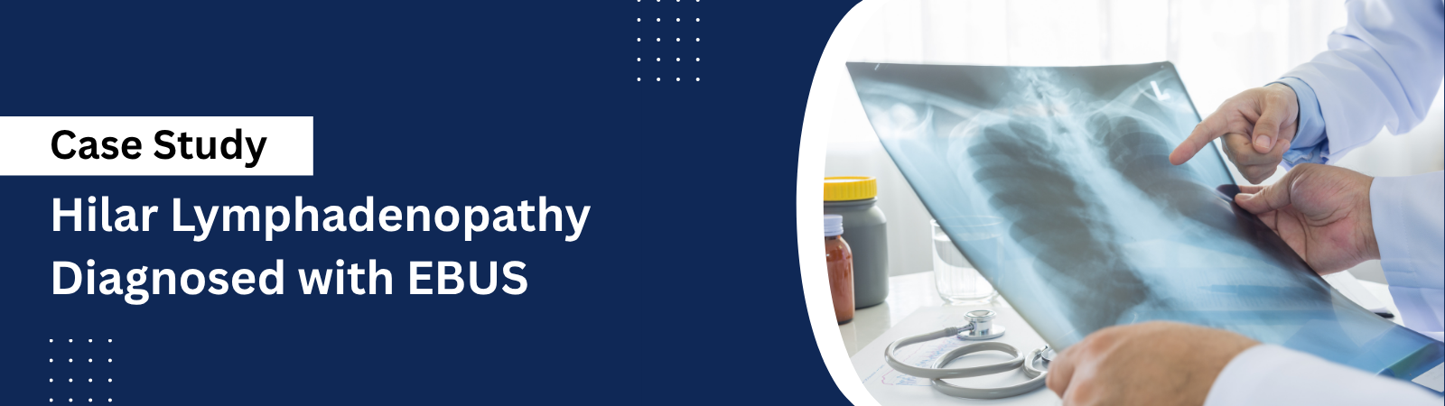
Bilateral Hilar Lymphadenopathy Diagnosed with EBUS-Guided Needle Aspiration
Patient Profile
| Age | 42 years |
| Gender | Female |
| City | Noida |
| Presenting complaint | Dry cough and mild breathlessness for three months, with fatigue |
| Imaging finding | Bilateral hilar lymphadenopathy on chest X-ray, confirmed on CT |
| Diagnosis | Sarcoidosis, confirmed on EBUS-guided transbronchial needle aspiration (EBUS-TBNA) |
| Procedure | EBUS-guided needle aspiration of mediastinal and hilar lymph nodes |
| Outcome | Definitive tissue diagnosis obtained without surgery. Treatment started. |
The patient’s identity is withheld in line with confidentiality guidelines. All other details are recorded from the clinical record.
The Problem
The patient was a 42-year-old woman with a three-month history of dry cough, mild breathlessness on exertion, and persistent fatigue. A routine chest X-ray showed enlargement of the lymph nodes at both lung hila, and she was referred to a pulmonologist in Noida for further evaluation.
A contrast CT of the chest confirmed bilateral hilar and mediastinal lymphadenopathy. Enlarged lymph nodes in this distribution have several possible causes, including sarcoidosis, tuberculosis, and lymphoma. These conditions require very different treatment, so a tissue diagnosis was essential before any therapy could begin.
The traditional route to these lymph nodes is mediastinoscopy, a surgical procedure performed under general anaesthesia through an incision at the base of the neck. It carries surgical risks, needs a hospital stay, and leaves a scar. The uncertainty of an unexplained abnormal scan, together with the prospect of chest surgery for a diagnosis, was a significant source of anxiety for the patient.
CONSULTATION & TREATMENT PLAN
What Was Assessed
- Detailed clinical history covering the duration of cough, breathlessness, fatigue, fever, night sweats, and weight loss.
- Review of the chest X-ray and contrast CT to map the size, number, and stations of the enlarged lymph nodes.
- Blood investigations, including serum ACE and calcium, and screening for tuberculosis.
- Assessment of the differential diagnosis: sarcoidosis, tuberculosis, and lymphoma.
- Evaluation of fitness for a bronchoscopic procedure under moderate sedation.
Why This Approach Was Chosen
Dr. Manu Madan recommended EBUS-guided transbronchial needle aspiration (EBUS-TBNA) as the first-line diagnostic procedure, in preference to open mediastinoscopy, for clear reasons:
- Minimally invasive: the lymph nodes are reached through the airway using a bronchoscope fitted with an ultrasound probe, with no incision and no scar.
- Real-time accuracy: the ultrasound shows the needle entering the node as the sample is taken, allowing precise targeting of multiple nodal stations in one session.
- Safer profile: EBUS avoids general anaesthesia and the surgical risks of mediastinoscopy, and is completed as a day-care procedure.
- Comparable diagnostic yield: for bilateral hilar lymphadenopathy, EBUS-TBNA achieves a diagnostic accuracy comparable to surgical biopsy in most cases.
Procedure Details
The procedure followed the standard protocol for EBUS treatment in Noida, performed under moderate sedation with continuous monitoring. The technique builds on the same airway access used in a conventional bronchoscopy, with the addition of an ultrasound probe at the tip of the scope.
Step by Step
- Pre-procedure assessment completed, fasting confirmed, and informed consent obtained.
- Moderate sedation administered, with continuous monitoring of oxygen levels and vital signs.
- EBUS bronchoscope passed through the mouth into the airways.
- Ultrasound used to visualise the enlarged mediastinal and hilar lymph nodes in real time.
- A fine needle passed through the bronchoscope into the targeted nodes under direct ultrasound guidance.
- Multiple aspirates taken from more than one nodal station to maximise diagnostic yield.
- Samples sent for cytology, histopathology, and microbiological testing, including for tuberculosis.
- Patient monitored in recovery for around two hours and discharged the same day.
Procedure Facts
| Procedure | EBUS-guided transbronchial needle aspiration (EBUS-TBNA) |
| Target | Bilateral hilar and mediastinal lymph nodes |
| Indication | Tissue diagnosis of bilateral hilar lymphadenopathy |
| Anaesthesia | Moderate sedation, as per institutional protocol |
| Approach | Through the airway; no incision |
| Intraoperative complications | None reported |
| Hospital stay | Day-care procedure; discharged the same day |
Post-Procedure Results
The aspirates showed non-caseating granulomas, and stains and cultures for tuberculosis were negative. Together with the clinical picture and blood results, this confirmed a diagnosis of sarcoidosis. A surgical biopsy was avoided entirely.
With a definitive diagnosis in hand, appropriate treatment was started without delay, and a monitoring plan was set for lung function and symptoms. The patient returned to her normal routine the day after the procedure.
Outcomes at a Glance
| Outcome | Result |
|---|---|
| Diagnostic yield | Definitive tissue diagnosis of sarcoidosis obtained at the first procedure. |
| Surgery avoided | No mediastinoscopy, no general anaesthesia, no incision, no scar. |
| Complications | None reported. |
| Hospital stay | Day-care; discharged the same day. |
| Treatment | Started promptly on the basis of a confirmed diagnosis. |
PATIENT FEEDBACK
Recorded during clinical follow-up:
“I had been told I might need chest surgery just to find out what was wrong, which was a difficult prospect. Dr. Manu Madan explained that the same answer could be obtained through the airway with EBUS. The procedure was completed in under an hour, I went home the same day, and within a week I had a confirmed diagnosis and a treatment plan.”
Post-Procedure Care and Recovery
Instructions Given to the Patient
- Avoid eating or drinking for two hours after the procedure until the sedation and throat numbness wear off.
- Expect a mild sore throat or a small amount of blood-streaked sputum for a day; report anything more than this.
- Avoid driving or operating machinery on the day of the procedure.
- Attend the review appointment for the biopsy results, usually available within three to seven days.
- Begin the prescribed treatment once the diagnosis is confirmed, and attend scheduled follow-up with lung function testing.
Recovery Timeline
| Timeframe | What the Patient Can Expect |
|---|---|
| Day of procedure | Two hours of monitored recovery, then discharge home. Mild throat discomfort is common. |
| Day 1 to 2 | Return to normal activities. Any throat soreness settles. |
| Day 3 to 7 | Pathology results available. Diagnosis discussed and treatment plan confirmed. |
| Week 2 onwards | Treatment underway with symptom monitoring. |
| Month 3 | Follow-up review with lung function testing to assess response to treatment. |
This case was managed by Dr. Manu Madan, Senior Consultant in Respiratory and Sleep Medicine at Medanta Hospital, Noida